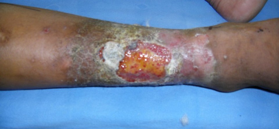
Venous Hypertension

SAMSUNG DIGITAL CAMERA
Leg ulcers are common and mostly due to venous hypertension. 40% of the venous ulcers are due to varicose veins. Surgical correction of varicose veins mostly leads to the healing of ulcers.
When the doctor clinically examines a leg ulcer he has 3 conditions in mind –
- Old DVT.
- Arterial disease.
- Varicose veins due to valvular insufficiency.
The diagnosis is made bed side by examining the patient and seeing the turgid the veins are. The hand held Doppler tells us about the reflux of blood while the patient is standing. And finally a colour Doppler study reveals the size of the veins and the reflux in the veins.
Venous hypertension affects over 2% of the population and is due to chronic venous insufficiency involving the deep and the superficial venous systems. In the deep system it could be due to post thrombotic syndrome and in the superficial system due to venous insufficiency.
The causes of chronic venous insufficiency (CVI) are:
1. Gravitational reflux – valvular dysfunction leads to venous insufficiency. This causes early pooling of venous blood after muscular contraction progressive and sustained increase in calf vein pressure. This is ambulatory venous hypertension causing capillary dilatation and leakage of plasma proteins. The compartmental pressures rise within the veins during muscle contraction. This is the hydraulic ram effect producing localized venous hypertension followed by oedema. The tissue pressure due to oedema continues to rise till it counteracts the high venous pressure and then stops pouring fluid into the tissue.2. Due to venous hypertension in a dependent limb the leukocutes accumulate. The hypoxic endothelial cells stimulate adherence of WBC and activate them which leads to release of O2 free radicles, collagenase and elastase which in turn injure the surrounding tissue.
Signs of venous hypertension – in order of occurrence
- Early sign – venous hypertension.
- Pigmentation.
- Lipodermatosclerosis.
- Ulceration.
Final diagnosis is based on Colour Doppler study which will show us:
- Venous reflux at LSV, SSV and perforators.
- If DVT is present in the leg veins.
- Flow augmentated by calf compression of calf, deep inspiration or valsalva manoeuvre.
Treatment of venous ulceration
- Elastic compression stockings.
- Surgery.