False/Pseudo Aneurysm
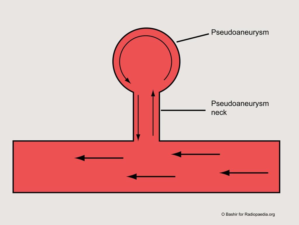
False aneurysms, also known as a pseudoaneurysm, is when there is a breach in the vessel wall such that blood leaks through the wall but is contained by the adventitia or surrounding perivascular soft tissue. A direct communication of blood flow exists between the vessel lumen and the aneurysm lumen through the hole in the vessel wall. The risk of rupture is higher than that of a true aneurysm of comparable size due to poor support of the aneurysm wall and thus false aneurysms generally require treatment.
Pathology
Aetiology
trauma (dissection or laceration)
iatrogenic (dissection, laceration or puncture), e.g. arterial catheterisation (accounts for most cases in this category 4 ), biopsy, surgery
spontaneous dissection
fibromuscular dysplasia (dissection)
mycotic aneurysm (inflammatory digestion of the vessel wall)
myocardial infarction (left ventricular false aneurysm)
regional inflammatory process
acute pancreatitis
chronic pancreatitis
vessel injury/erosion due to a tumour: relatively uncommon
vasculitides
Behcet syndrome
giant cell arteritis
Takayasu arteritis
systemic lupus erythematosus
polyarteritis nodosa
penetrating atherosclerotic ulcer
Location
They can involve any arterial segment or even a cardiac chamber. Examples include
aortic pseudoaneurysm: traumatic aortic pseudoaneurysm
femoral artery pseudoaneurysm: relatively common site due to femoral punctures
carotid artery pseudoaneurysm
visceral arterial pseudoaneurysm
hepatic arterial pseudoaneurysm
gastroduodenal arterial pseudoaneurysm
splenic arterial pseudoaneurysm
prei-pancreatic pseudoaneurysm
renal arterial pseudoaneurysm
peripheral arterial (limb) pseudoaneurysm
left ventricular pseudoaneurysm
brachiocephalic artery pseudoaneurysm
Radiographic features
Some of the imaging features may be dependent on location.
Ultrasound
Due to the turbulent forward and backward flow, a characteristic yin-yang sign may be seen on colour flow while a "to and fro" pattern may be seen with pulsed Doppler.
CT
Hypoattenuating (non-contrast) or hyperattenuating contrast-enhanced) smooth walled sac adjacent to an artery, usually with a communication.
Endovenous Laser Treatment
Endovenous Laser Treatment for the elimination of varicose veins is quickly becoming the gold-standard in the treatment of varicose veins. Endovenous Laser Treatment uses laser energy, which is simply a highly concentrated beam of light. Medical lasers work by delivering this light energy to the targeted tissue with extreme precision, so as not to affect the surrounding tissue. Lasers have proven their safety and effectiveness through years of use in all types of medical procedures, from eye surgery to dermatology. In the hands of a skilled physician, lasers offer far less risk for complications than conventional surgery.
In endovenous laser treatment, a thin fiber is inserted into the damaged vein through a very small entry point in the skin. A laser light is emitted through the fiber, as the fiber is pulled back through the vein, it delivers just the right amount of energy. The targeted tissue reacts with the light energy, causing the vein to close and seal shut. The veins that are closed are superficial veins that handle less than five percent of the body's blood flow. The blood is automatically routed to other, healthy veins.
Some physicians are now using a jacketed fiber, which prevents any contact between the fiber and the vein wall. This prevents much of the pain and bruising that is often associated with the more conventional method of ligation and stripping. Some patients may experience temporary soreness or some slight swelling, which can be treated effectively with over-the-counter, non-aspirin pain relievers and typically subsides within the first five days.
The procedure is minimally invasive and requires no general anesthesia. Only local anesthetic is used to numb the area where the physician is working. Patients are encouraged to walk immediately after the procedure and can resume normal activities the same day.
Recovery
The Endovenous Laser Treatment procedure is designed to make sure there is minimal disturbance to the patient’s life and activities immediately following. There is no recovery time after this procedure for the treatment of painful varicose veins. You may feel slight tightening of the area and some mild bruising can occur, but this is normal and is not painful. After the procedure, there is no recovery time, but we do encourage that no heavy lifting be done afterward. We also encourage minimizing vigorous exercise; depending on each patient this will vary.
Result
Once the varicose vein is closed by the Endovenous Laser Treatment procedure, the blood is then rerouted to deeper veins and circulation improves significantly. After a short time, normal exercise and physical activity will increase and be much less painful. Prior to the treatment of varicose veins with the EVLT, patients experienced high levels of discomfort in small movements. Walking that was once difficult will now be enjoyable after the laser treatment. Not only will your circulation improve thereby improving your overall health, but also the appearance of unsightly varicose veins is significantly minimized. Varicose veins cause embarrassment, and attempts to conceal them are difficult. After this minimally invasive procedure is completed there’s no need to worry about showing off some leg, uncovering your hands and other areas.